

When does a posterior approach make sense?
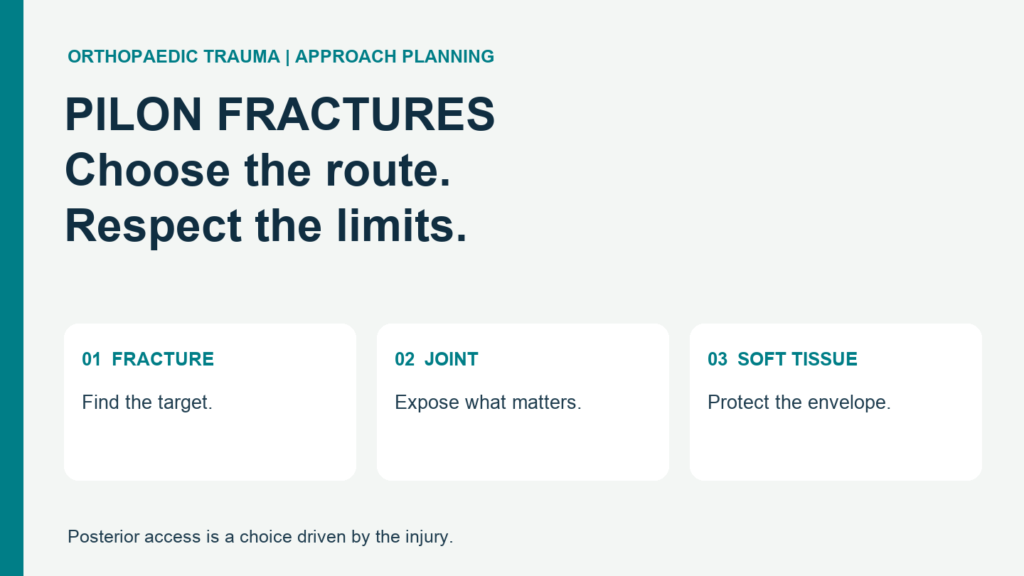
A posterior approach to pilon fractures may help when the back of the distal tibia is short, displaced, or crushed at the joint. It is a route chosen for a problem. It is not a badge of advanced care. A pilon fracture breaks the weight-bearing end of the shinbone at the ankle. The task is to restore the joint, support the bone, and protect the skin. The 2026 review by Tiee, Stewart and Usmani links route choice to the site of injury, the planned fixation, and soft tissue. It also notes limited data to guide the choice among rear approaches. That matters. A neat incision cannot rescue a poor plan. Ask what the chosen route lets the team see and fix. Then ask what remains hidden. Those two answers should drive the choice.
What must the CT-based plan answer?
Map the fragments before naming the incision. The supplied review puts five issues at the heart of the choice: fracture shape, joint impaction, trapped soft tissue, fibula injury, and skin condition. A posterior approach to pilon fractures should address that map. Look for the back fragment and any split that runs toward the inner ankle. Check the middle of the joint, too. A small piece driven into the bone can block a sound reduction. “Die-punch” describes this impacted piece; it is not just another loose chip. The plan must explain how the team will reach it. Keep this short checklsit in view:
- Bone: where are the main breaks and gaps?
- Joint: what is crushed, tilted, or trapped?
- Soft tissue: where can the skin tolerate access?
A scan is useful when its findings change the plan. Naming fragments alone is not enough.
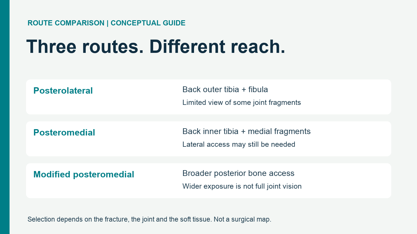
Posterolateral access: useful, but not a full joint view
The posterolateral route can reach the back outer tibia and the fibula through one skin incision. That is a clear benefit when both need work. AO Surgery Reference describes its use for selected rear-dominant patterns, especially with little front surface involvement. The tibial corridor lies between the peroneal tissues and flexor hallucis longus (FHL). The sural nerve and nearby vessels need protection. Access, however, is not the same as sight. Small joint pieces may remain hard to see. A posterior approach to pilon fractures must account for that limit before fixation locks the scene in place. If the break extends toward the inner side, this route may leave the key problem out of reach. One incision sounds efficient. It earns that label only when it gives the access the fracture actually needs.
Posteromedial access: match it to the medial fragment
The posteromedial route targets the back inner tibia and selected medial fragments. It can support direct reduction and the placement of a posterior buttress plate. That plate helps resist the force that would displace the fragment again. The AO guide to posteromedial exposure describes several deep intervals; the fracture location guides which one is used. The tibial nerve and posterior tibial vessels make this a demanding choice. Protection is part of the route, not an optional extra. The supplied article also highlights trapped tendons or other soft tissue as reasons to assess medial access. A posterior approach to pilon fractures should not be chosen on bone shape alone. What lies between the pieces may matter just as much. This route does not automatically solve a far lateral break or a fibula fracture. Separate access may still be needed.
Does a modified posteromedial route solve everything?
A wider rear exposure does not guarantee a better view of the whole joint. The supplied article describes an extended, modified posteromedial route for broader access across the back of the tibia. It cites Assal and colleagues’ 2014 technique report, which combined a modified rear route with anterior access for complex injuries. The distinction matters: broader bone exposure and complete joint inspection are different goals. The source also reports cadaveric exposure figures. These are measurements of access under study conditions, not patient success rates. They should never become a claim that one incision is “91% effective.” A posterior approach to pilon fractures still needs a plan for hidden joint damage and nerve protection. Extending a route is a response to a specific need. More exposure, by itself, is not a treatment goal. The fracture sets the scope.
When should posterior fixation happen?
The soft tissue sets the pace; a fixed timetable cannot. The supplied review describes early, limited rear fixation with a spanning external fixator as one option in selected cases. A restored back column can then serve as a guide for later reconstruction. This is a strategy, not a rule for every injury. A posterior approach to pilon fractures must fit the whole sequence, including any front incision and later screws. A plate placed early should not block the next step. AO’s approach-planning guidance stresses planning future incisions during initial care and allowing injured soft tissue to recover before definitive fixation when needed. It also notes that most pilon fractures are best approached from the front. Keep that context. A strong rear fixation plan can complement front access. It does not erase the need for it.
Which route is best? Demand a fracture-specific answer
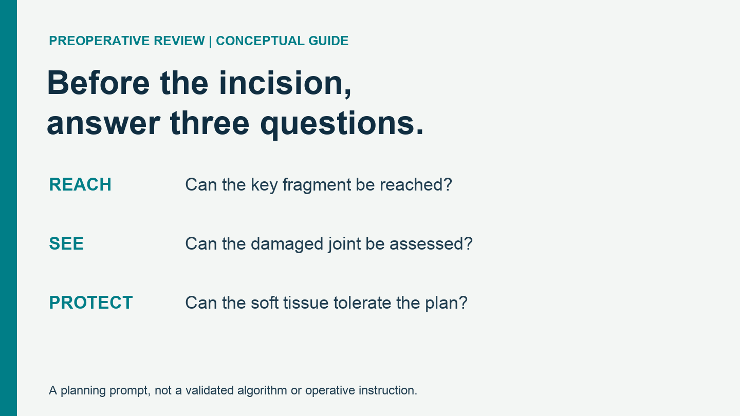
No single incision wins every case. The best posterior approach to pilon fractures is the one that meets the actual goals with acceptable tissue risk. This is the practical reading of the supplied review, not a validated scoring tool. Start with three questions:
- Can the team reach the displaced fragment?
- Can it assess and restore the damaged joint surface?
- Can fixation support the bone without sacrificing the skin?
If an answer is unclear, the plan needs work. A second route may be needed. So may a different sequence. Neither is a failure of technique. The failure is to force a complex injury into a favoured incision. For surgeons and trauma teams, the useful comparison is access, limits, and tissue cost. For patients, ask why the route fits your scan. No diagram or general article can choose your operation.