

What is bone transport, and what can it actually fix?
Bone transport moves a living segment of bone across a gap while new bone forms behind it. It is one way to rebuild a limb after major bone loss, often with an external frame. The idea is simple. The treatment is demanding. A surgeon makes a controlled cut in healthy bone, then guides the segment toward the far end of the defect. The new tissue must mature, and the moving segment must unite with the bone it reaches. These are two separate healing tasks. Closing a gap on an X-ray does not prove either is complete. This guide explains the process, key risks and questions to ask before treatment. It is general education, not a schedule for adjusting a frame or deciding how much weight to put through an injured limb.
How does new bone grow across a defect?
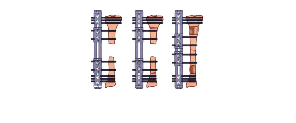
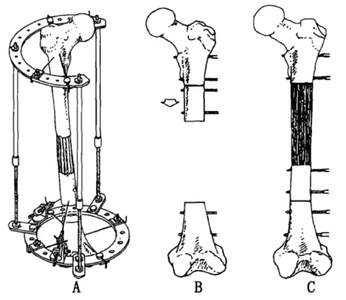
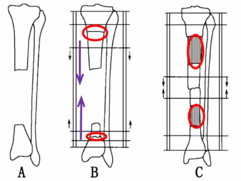
The key process is distraction osteogenesis: slow, controlled separation of a surgically divided bone can stimulate new bone in the gap. Think of the moving segment as a bridge builder that leaves a path behind it. That path begins as living repair tissue, not hard bone ready for full load. It needs blood supply, stable support and time to gain strength. At the other end, the segment reaches the docking site, where two bone surfaces must join. Bone transport therefore asks the team to watch both the new bone column and the contact point. The source diagrams show how one or more moving segments can fill a defect. They explain the mechanics; they cannot predict a patient’s healing speed, safe load or final limb function.
Who may be considered for this treatment?
A large gap after trauma or removal of infected bone can make simple fracture repair inadequate. In selected patients, bone transport can help rebuild missing length and address a difficult nonunion. Yet a large defect alone does not settle the choice. The state of the skin, muscle, blood vessels, nerves and nearby joints matters too. So does the person’s ability to cope with a long course of care. Infection control and a viable soft-tissue envelope are central to the plan. For infected defects, this surgical reference on infection and skeletal reconstruction explains the role of debridement, suitable antibiotics and stable fixation. Moving bone is not a substitute for those steps. Ask what problem each stage will solve, which findings could change the plan, and why the proposed approach suits this limb.
How long does bone transport take?
There is no honest one-line recovery date. Treatment includes surgery, a rest period before movement, gradual transport, bone maturation and rehabilitation. The often quoted figure of about 1 mm per day describes a commonly used movement rate, not a promise and not a home instruction. The surgeon adjusts timing and rate to the patient’s response. A simple example shows the trap: moving one segment across a 50 mm gap at 1 mm per day would take about 50 days of movement. That calculation excludes pauses, initial healing, docking and the time needed for the new bone to become strong. It is not the total time in a frame. A useful bone transport plan should explain each phase, review dates and the evidence required before support is reduced or removed.
One moving segment or two: what is the difference?
The original article illustrates both a single segment moving across a defect and two segments advancing toward a central gap. In the second arrangement, new bone develops at two distraction sites. That can change how far each segment must travel, but it does not erase the need for healing at the contact point. Nor does a more complex drawing prove a better result. Frame layout must follow the defect and the living tissues around it. Segment size, fixation points, alignment and joint movement all need a workable plan. Bone transport is a reconstruction strategy, not a contest to add more hardware. When reviewing the diagrams, ask where the new bone will form, where docking will occur, and how the team will detect drift or poor progress before it becomes a larger problem.
What can go wrong during recovery?
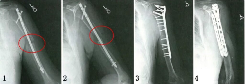
The risks deserve space before the success stories. Pin-site infection, joint stiffness, poor regenerate formation and docking-site nonunion can disrupt the course of care. A frame may need adjustment, and further surgery may be required. Even after removal, weak new bone can fracture. These concerns are described in clinical research on regenerate and docking-site healing; they should be part of consent, not a footnote. Bone transport follow-up must assess comfort, skin and pin sites, limb alignment, joint motion and serial images. Do not treat a new problem as the price of being brave. Ask the treating team which symptoms need an urgent call and whom to contact outside clinic hours. A written care plan is more useful than a vague instruction to return if things get worse.
What do published results really tell us?
A successful case shows that a method can work. It does not tell you that the same outcome will occur in every limb. One retrospective study of 12 infected forearm defects reported infection-free union in all included patients and a mean external fixation time of 232.6 days. That is a small, selected group, not a universal timetable or success rate. The study also used staged infection treatment and close follow-up. Read the patient selection and treatment burden alongside the result. Bone transport outcomes should be judged by pain, function, infection control and the need for extra procedures, as well as union on an image. A tibial defect and a forearm defect face different loads and functional goals. Mixing them into a single attractive percentage hides the details that matter.
What should you ask before choosing a reconstruction plan?
Start with the goal: a healed, useful limb and a treatment burden the patient can realistically manage. Ask the limb reconstruction team to compare bone transport with the alternatives that fit the actual defect. Depending on the case, discussion may include graft-based reconstruction, an induced membrane approach or vascularized bone transfer. Each has trade-offs; none can be ranked from a photograph alone. Bring prior operation notes, imaging and infection results to the consultation. Then ask for a clear explanation of the likely stages, possible extra procedures and follow-up demands. The next step is an individual assessment, not choosing a frame from a picture. The questions below can keep that discussion concrete. A good plan should explain what progress looks like and what the team will do if healing stalls.
- What must be done to control infection and protect soft tissue?
- How will new bone and docking-site union be checked separately?
- What are the rules for pin care, exercise and weight bearing?
- Which costs, clinic visits and further operations should be planned for?
Source and illustration notes
Adapted from the supplied Chinese educational article on bone transport, dated 18 September 2026. Diagrams are reproduced from that source for explanation and do not represent a treatment prescription or a promised outcome. Original article and illustration context. Supporting clinical sources are linked above. Last reviewed: 18 September 2026.