In radiographic imaging, the patient’s positioning plays a pivotal role in determining the quality of the image and the diagnostic accuracy. Due to the overlapping nature of anatomical structures, achieving a clear image can be challenging. To overcome this, radiologic technologists must master various positioning techniques and X-ray projection methods to isolate specific body parts and provide a more accurate view. This article explores the critical relationship between X-ray projection methods and patient positioning, offering insights into how proper techniques can optimize image quality.
Key Terminology: Understanding Patient Positioning and Projections
When discussing X-ray projections, it’s essential to be familiar with basic terms that help technologists position patients accurately for optimal imaging.
- Anterior (front) vs. Posterior (back): Refers to the front and back of the body, respectively.
- Superior (top) vs. Inferior (bottom): Describes the upper and lower parts of the body.
- Medial (toward midline) vs. Lateral (away from midline): Indicates proximity to the body’s centerline.
- Proximal (closer to the body center) vs. Distal (farther from the body center): Used to describe limb positioning.
- Cranial (toward the head) vs. Caudal (toward the feet): Refers to directional terms based on body orientation.
These terms are crucial when describing patient positioning for radiographic exams, ensuring that the correct anatomical structures are captured.
X-ray Projections: How Positioning Affects Image Quality
X-ray projection methods describe the path the X-ray beam takes through the body. Different projections reduce overlap and provide clearer anatomical details, helping to improve diagnostic accuracy.
- Anterior-Posterior (AP) Projection: The X-ray beam enters from the front (anterior) and exits from the back (posterior). This is commonly used for limb and pelvic imaging.
- Posterior-Anterior (PA) Projection: The beam enters from the back and exits from the front, often used in chest X-rays to minimize heart magnification.
- Lateral Projection: The beam passes from one side of the body to the other, ideal for imaging the chest or joints.
- Oblique Projection: The beam passes through the body at an angle, useful for complex structures like the spine or wrist.
- Axial Projection: The beam travels along the body’s long axis, commonly used for head and neck imaging.
By utilizing these different projection methods, radiologic technologists can significantly enhance image clarity and diagnostic precision.
Common X-ray Exams and Positioning Requirements
Different X-ray exams require specific patient positioning to ensure the best possible images. Below are some common exams and their positioning guidelines:
- Hand X-ray: The patient sits with the elbow bent and the hand placed palm down on the image receptor. For an oblique view, the hand is rotated approximately 45 degrees.
- Wrist X-ray: The wrist is placed flat on the image receptor with the hand pronated. In lateral views, the wrist is aligned horizontally with the forearm.
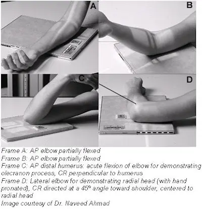
- Elbow X-ray: For the AP view, the elbow is extended with the hand supinated. A lateral view requires the elbow to be bent at a 90-degree angle.
These standardized positioning protocols ensure that the resulting images are clear and suitable for accurate diagnosis.
Clinical Significance: How Proper Positioning Impacts Diagnosis
In clinical practice, X-ray imaging is one of the most frequently used diagnostic tools. Correct patient positioning not only improves image quality but also helps avoid misdiagnoses.
- Chest X-rays: Frontal and lateral views allow the radiologist to assess heart size, identify pneumonia, and evaluate pneumothorax volume.
- Abdominal X-rays: Upright and supine views help detect free air, bowel obstructions, and abnormal calcifications.
- Mammography: Proper positioning is crucial for detecting microcalcifications, a key indicator of potential malignancy.
Thus, standardized positioning techniques are essential for obtaining high-quality images and ensuring accurate diagnoses.

Conclusion
The relationship between X-ray projection methods and patient positioning directly impacts the quality of the resulting images and the accuracy of the diagnosis. Correct positioning not only optimizes the image but also helps avoid diagnostic errors. Radiologic technologists play a vital role in this process by ensuring precise positioning and utilizing the appropriate projection techniques to deliver the best possible images for physicians.
For more detailed information on X-ray projection methods and patient positioning, visit RadiologyInfo, a trusted source for radiology knowledge.
Meta Description: Discover the crucial relationship between X-ray projection methods and patient positioning, and how proper techniques can optimize image quality and diagnostic accuracy.
Disclaimer:
This article and all articles on this website are for reference only by medical professionals; specific medical problems should be treated promptly. To ensure “originality” and improve delivery efficiency, some articles on this website are AI-generated and machine-translated, which may be inappropriate or even wrong. Please refer to the original English text or leave a message if necessary. Copyright belongs to the original author. If your rights are violated, please contact the backstage to delete them. If you have any questions, please leave a message through the backstage, or leave a message below this article. Thank you!
Like and share, your hands will be left with the fragrance!


