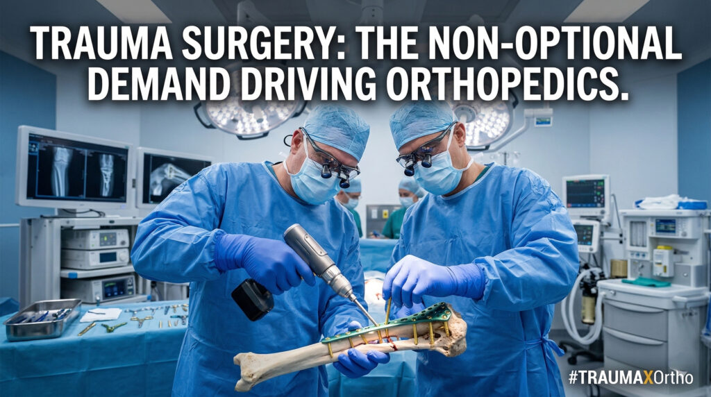


Open reduction and internal fixation of fractures are the most common and basic operations for orthopedic surgeons. The purpose of surgical treatment of fractures is to restore normal anatomy (or functional reduction) as much as possible to create the hardware prerequisite for functional recovery. Fracture reduction is the most important part. Usually, fracture reduction is based on normal anatomical angles or the restoration of local complete morphology. However, bone development varies from person to person. The most accurate reduction standard is based on the patient’s normal anatomy. Therefore, the anatomy of the healthy side is an excellent reference standard. Foreign scholars have proposed a fracture reduction assessment method based on “healthy side limb imaging films”, which is widely used in the internal fixation of fractures in various parts of the clinic, such as tibia and fibula, ankle, distal radius, etc.
The principles and steps of this method are as follows:
- X-ray of the anteroposterior and lateral positions of the healthy limb (based on the fluoroscopic position of clinical evaluation);
- Trace the bone contour of the healthy side image on “transparent film” or white paper, and place the healthy side image on the C-arm and the left side of the screen;
- Flip the traced image, which is the standard for the reduction of the affected side, and use it as the standard for surgical reduction.
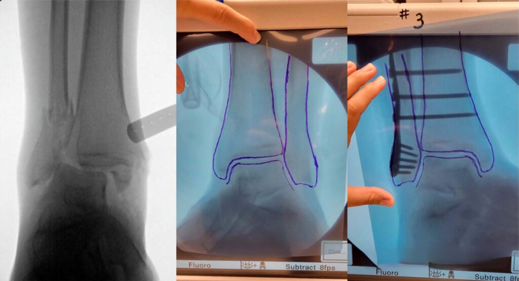
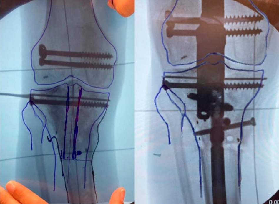
#Ankle
As shown in the figure below, first perform normal X-ray anteroposterior and lateral views of the healthy side. When performing an X-ray of the healthy side, the intraoperative X-ray position of the affected side must be considered. If it needs to be elevated or rotated during the operation, the same position change must be performed when performing an X-ray of the healthy side.
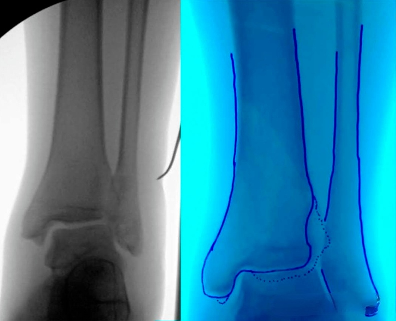
After fluoroscopy, use transparent film to trace the bone contour of the healthy side, and then flip it over to obtain the reduction standard of the affected side (as shown below):
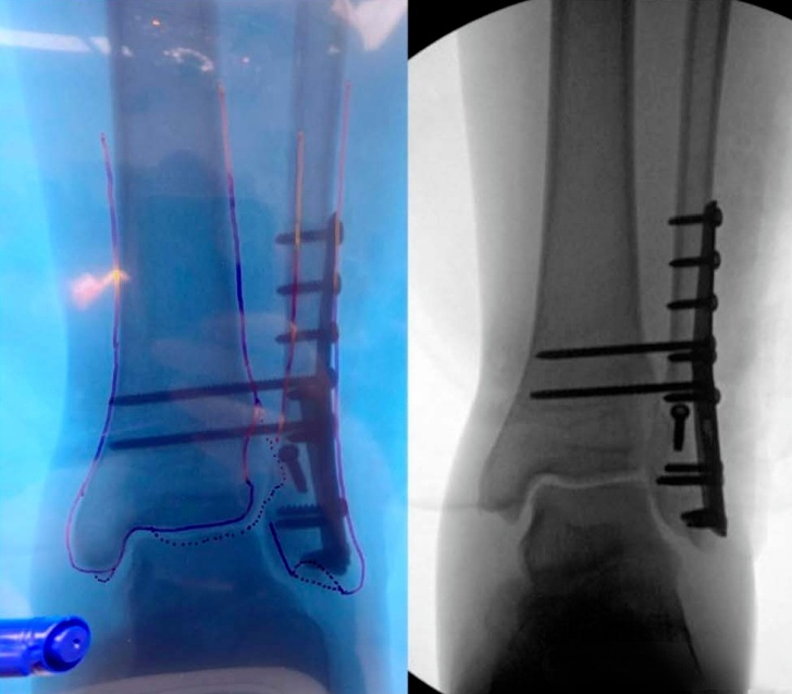
Another case of pronation-abduction ankle fracture also used this method:
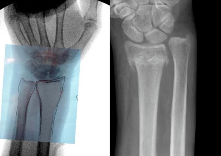
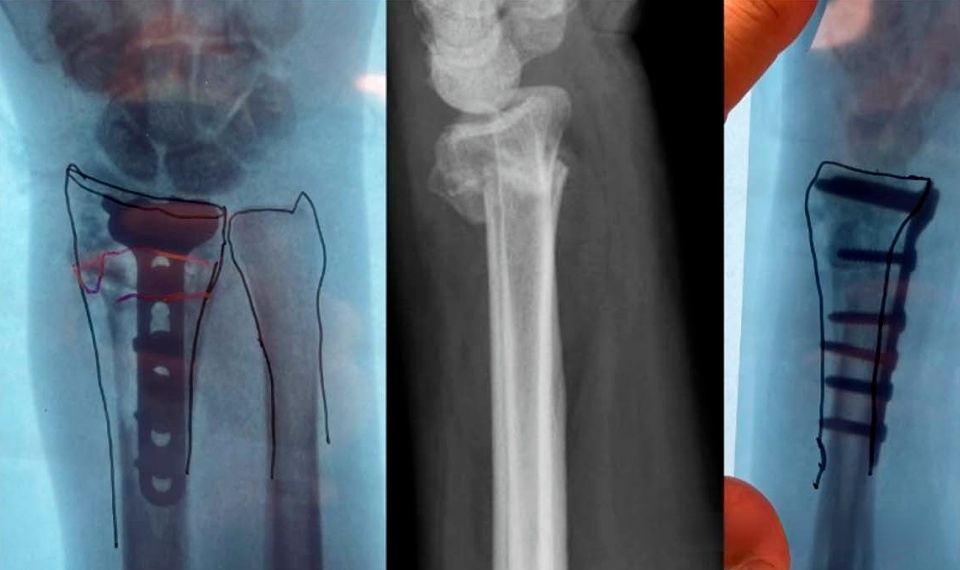
#Distal radius
Similar to the ankle joint, the reduction of distal radius fractures involves palmar tilt, ulnar deviation, and height restoration. The above parameters vary in the normal population. The most reliable reduction standard can be achieved by taking the unaffected wrist as a reference:
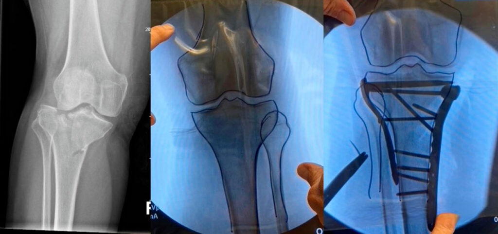
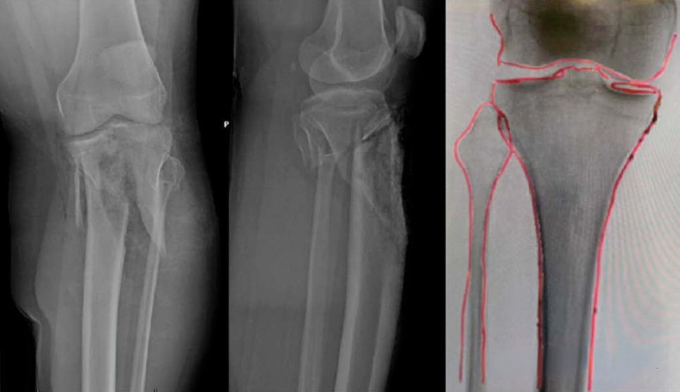
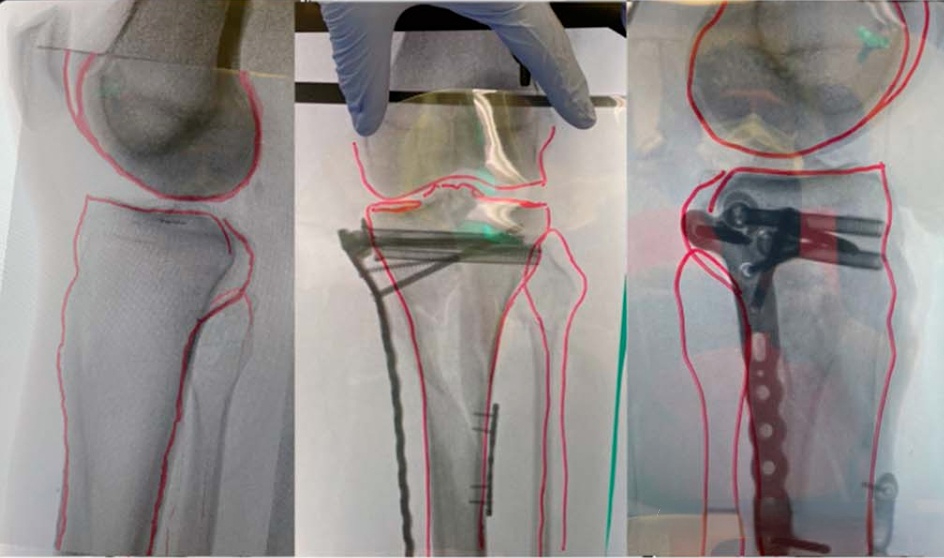
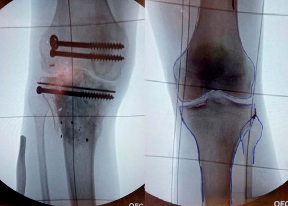
#Tibial plateau
The reduction of complex tibial plateau fractures is indeed difficult in clinical practice, especially for bicondylar fractures, which have collapsed and displaced joints and cannot be used as a reference like local articular surface fractures. Therefore, intraoperative reduction evaluation is difficult:
#Osteotomy
This method is more suitable for osteotomy correction of lower limb deformity:
This method is theoretically feasible, but in clinical practice, there are uncertainties in how to ensure that the healthy side and the affected side are in the same fluoroscopic position, and how to control the rotation of the limbs and the height of the C-arm tube. For reference by clinicians.
[Statement]: The concepts, technologies, and principles shared on this platform are all publicly available journals, published books, or online platform materials. The copyright belongs to the original author. The platform only organizes, summarizes, and shares them for learning reference. This platform is not responsible for the authenticity of the content and the effectiveness of the technology. The related medical behaviors generated based on the content pushed by this platform have nothing to do with the platform. Please choose carefully. If there is any infringement, please contact us to delete it.
Welcome to share, forward, and like the article in the lower right corner!