


Hot Topics and Controversies in Midshaft Clavicle Fractures
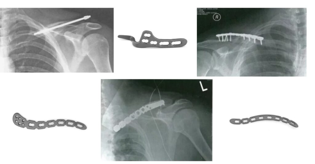
Clavicle fractures are among the most common types of fractures, accounting for 2.6% to 12% of all fractures. Of these, midshaft clavicle fractures make up a significant 80%, making it a key focus of research and treatment. Clavicle fractures not only impact bone structure but can also affect the function of the shoulder girdle, which […]
Research hotspots and progress of orthopedic implants
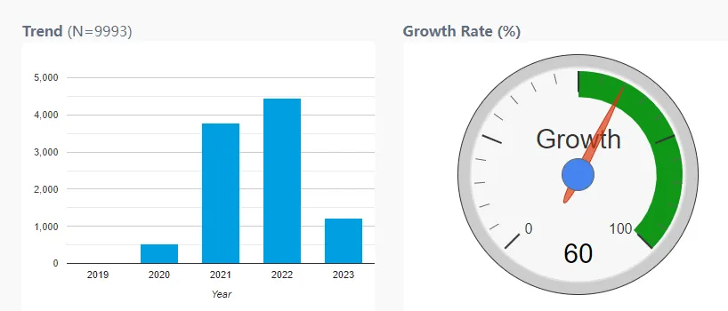
The repair and replacement of large-scale bone defects caused by diseases, trauma, and aging has been an important topic that humans have been studying for centuries. However, to date, the clinical treatment of large-scale bone defects remains a world problem. The use of orthopedic implants to reconstruct the structure and function of bone tissue in […]
Epiphysisitis and osteochondrosis: two common causes of bone pain in growing children
As a young orthopedic doctor, understanding the nuances of pediatric musculoskeletal disorders is crucial. This article introduces two common causes of pain in growing bones: apophysitis and osteochondrosis. While both conditions affect developing skeletal systems, they have distinct etiologies, presentations, and management approaches. Apophysitis: Traction-Induced Pain Apophysitis results from traction injury to the cartilage and […]
Causes and countermeasures of fracture nonunion: analysis from several papers
Causes and Treatment of Nonunion of Fractures Nonunion of fractures, also known as pseudoarthrosis, occurs when a fracture fails to heal without further intervention. This condition can persist regardless of how much time has passed since the initial injury. Understanding the causes of nonunion is critical for effective management and treatment. Causes of Nonunion of […]
Femoral Neck Dynamic Cross Nail System Product Introduction
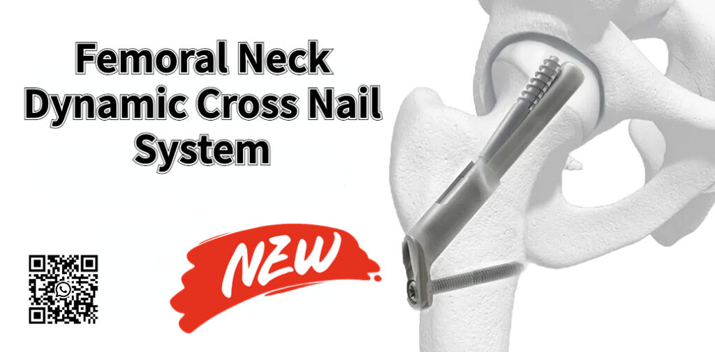
IndicationsFemoral neck fracture (AO type 31-B)(Intertrochanteric fracture and subtrochanteric fracture are contraindicated) Product Advantage 1Excellent angular stabilityThe main screw and the anti-rotation screw maintain overall angular stability with the locking screw, which can effectively prevent varus collapse and femoral neck shortening, and can provide more adequate angular stability compared to ordinary hollow screw treatment. Product […]
Vascular injury in orthopedic surgery: a comprehensive review of risks and prevention strategies
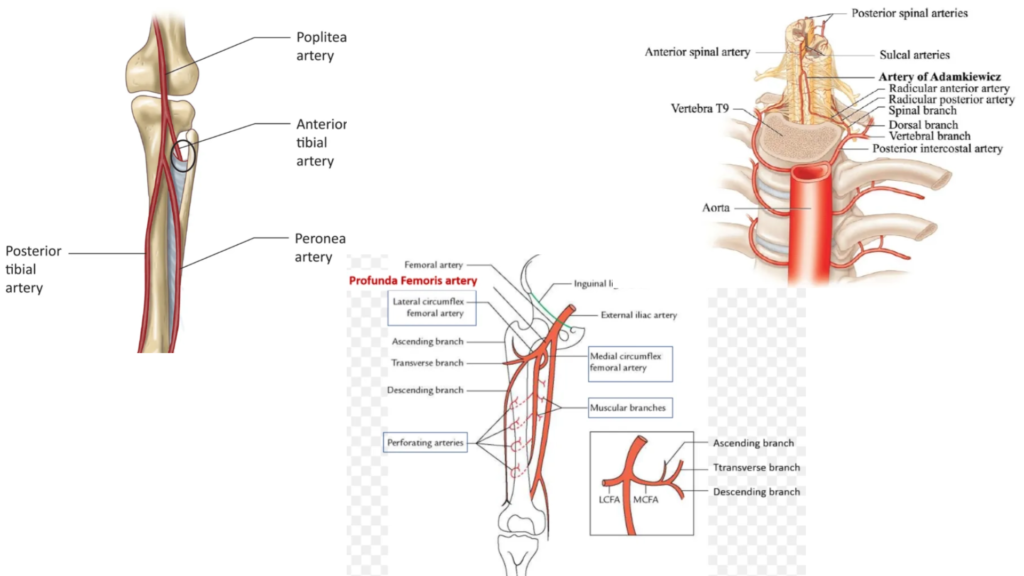
Orthopedic surgery, though transformative for patients suffering from musculoskeletal disorders, carries an inherent risk of vascular injury. While relatively uncommon, these complications can have devastating consequences, including limb loss and even death. This article provides a detailed review of the types of orthopedic surgeries most susceptible to vascular injury, the mechanisms underlying these injuries, and […]
Beware of vascular injury in orthopedic trauma: diagnosis and treatment

Vascular injury in orthopaedic trauma References: Disclaimer:This article and all articles on this website are for reference only by medical professionals; specific medical problems should be treated promptly. To ensure “originality” and improve delivery efficiency, some articles on this website are AI-generated and machine-translated, which may be inappropriate or even wrong. Please refer to the […]
Ten aspects of failed internal fixation of femoral shaft fractures: detailed causes and prevention
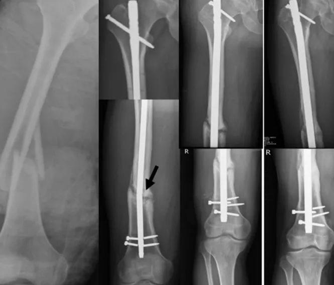
Today, let’s delve deeper into the causes and preventative measures for internal fixation failure in femoral shaft fractures: 1. Patient Factors: 2. Cerclage Wire Use:2. 3. Cerclage Wire Placement: 4. Screw Placement in Intramedullary Nailing (IMF): 5. Screw Placement in Plate Osteosynthesis (PO): 6. Choice of Implant and Fixation: 7. Technical Errors During Surgery: 8. […]
Analysis and selection of advantages and disadvantages of various treatment methods for multi-segmental tibial fractures
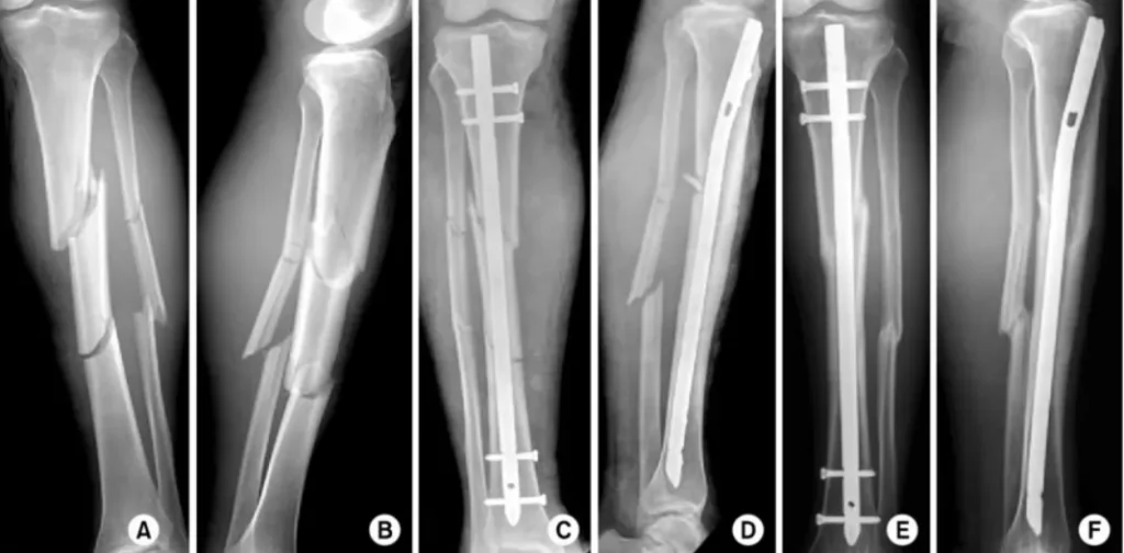
Segmental tibial fractures are complex injuries involving two or more distinct fracture lines and a free-floating middle fragment. This complexity demands a multidisciplinary approach and careful consideration of various factors when deciding on the optimal treatment. Here’s a breakdown of common treatment methods and their pros and cons: 1. Non-operative Treatment: 2. Surgical Treatment: a) […]
Be careful! There are these dangerous points in the internal fixation surgery of proximal humeral fracture
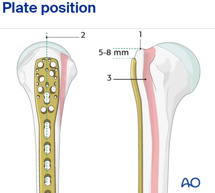
Proximal humeral fractures are very common in orthopedic clinical practice. This article will discuss several major pitfalls that need to be noted in open reduction and internal fixation and intramedullary nailing of proximal humeral fractures, aiming to provide guidance for young orthopedic surgeons to optimize surgical outcomes and reduce postoperative complications. Open reduction and internal […]