


Probability of Radial Nerve Recovery After Humeral Fracture Surgery: A Bayesian Insight
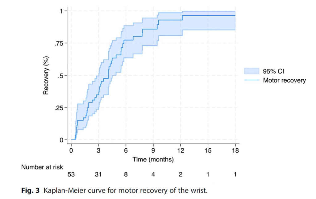
Meta Description: Discover the real-world probability of radial nerve recovery after humeral shaft fracture surgery. Learn how Bayesian analysis reveals recovery timelines, influencing surgical decision-making and patient communication. Understanding Radial Nerve Injury in Humeral Fractures Radial nerve palsy is one of the most concerning complications following humeral shaft fracture surgery. For decades, surgeons have debated […]
How to Insert Thoracolumbar Pedicle Screws? Let AO Principles Guide You
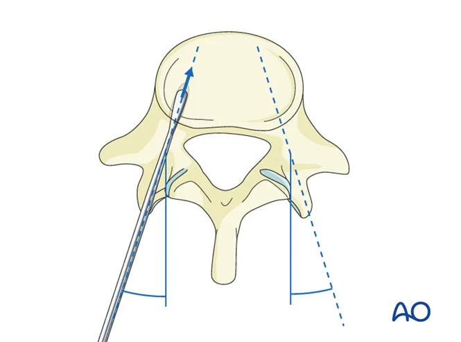
Meta Title: Thoracolumbar Pedicle Screw Insertion Guide – Step-by-Step AO Technique Meta Description: Learn how to insert thoracolumbar pedicle screws accurately and safely with this AO-based 7-step guide. Covers entry point location, angle control, and intraoperative risks. Why Pedicle Screw Insertion Matters in Spine Surgery Pedicle screw fixation is the cornerstone of spinal surgery for […]
Galeazzi Fractures: Step-by-Step Treatment Algorithm for Optimal Outcomes
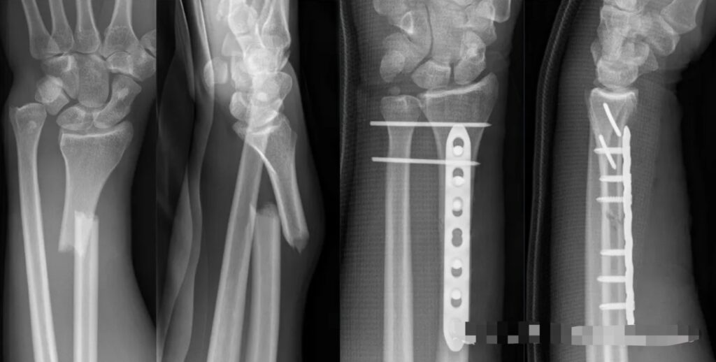
Galeazzi fractures, characterized by a fracture of the radial shaft with disruption of the distal radioulnar joint (DRUJ), are notoriously unstable injuries that demand precise surgical management. Without proper treatment, these fractures can result in chronic instability, pain, and functional impairment. This article provides a comprehensive treatment algorithm that ensures anatomic reduction, DRUJ stability, and […]
Distal Humeral Fractures: Intramedullary Nail vs Plate Fixation – Mechanics, Techniques, and Complications

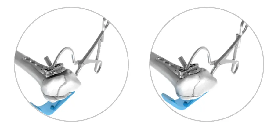
Distal humeral fractures represent a unique challenge in orthopedic trauma surgery. While less common than fractures of the humeral shaft or proximal humerus, their complex anatomy, variable fracture patterns, and propensity for complications demand a tailored approach. The debate between intramedullary nailing (IMN) and plate fixation (ORIF) continues to evolve, with both methods offering distinct […]
Exploring Advanced Techniques and Challenges in Orthopedic Trauma Surgery
Orthopedic trauma surgery is a constantly evolving field, where precision, innovation, and a deep understanding of biomechanics and biology are essential for success. From helical plates to minimally invasive osteosynthesis (MIO), this article delves into cutting-edge techniques, challenges, and solutions for fracture fixation. Whether you’re addressing implant fatigue, trochanteric fractures, or surgical invasiveness, these insights […]
Challenges, Biomechanical Considerations, and Treatment Choices in Fracture Fixation
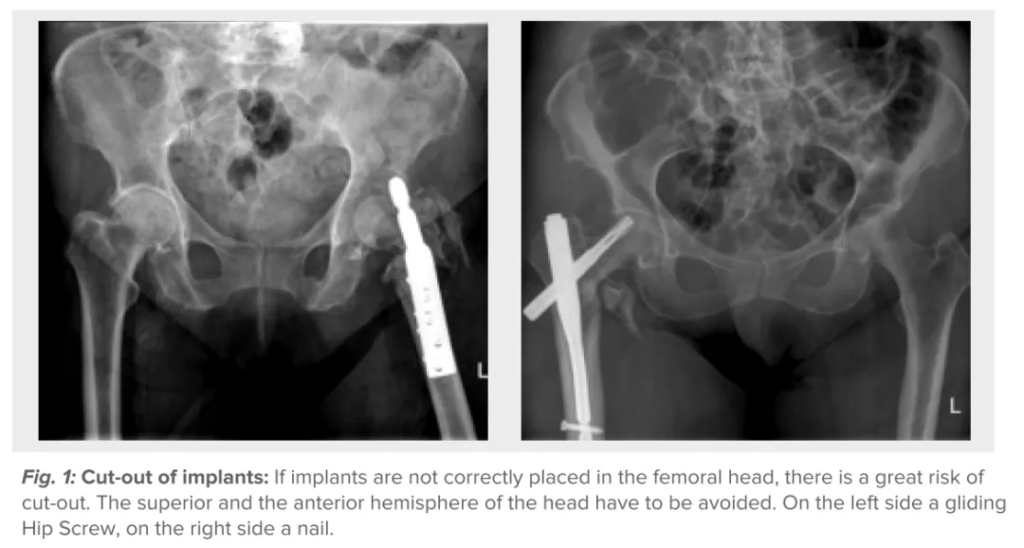
Fracture fixation remains one of the most complex areas of orthopedic surgery, demanding precision, technical expertise, and a deep understanding of biomechanics and biology. This article explores the challenges, biological and biomechanical considerations, and optimal treatment strategies for managing fractures, drawing insights from ICUC App case studies and clinical scenarios. Challenges in Fracture Fixation Fracture […]
Understanding Screws in Fracture Surgery: A Comprehensive Guide
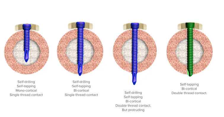
When it comes to fracture surgery, the choice of screws can make or break the success of the procedure. With multiple types of screws available, each offering unique properties, understanding their applications and limitations is critical for achieving optimal outcomes. This article explores the different types of surgical screws, their functions, advantages, and potential pitfalls, […]
Corona Mortis in Pelvic Fracture Surgery: Identification and Hemorrhage Management
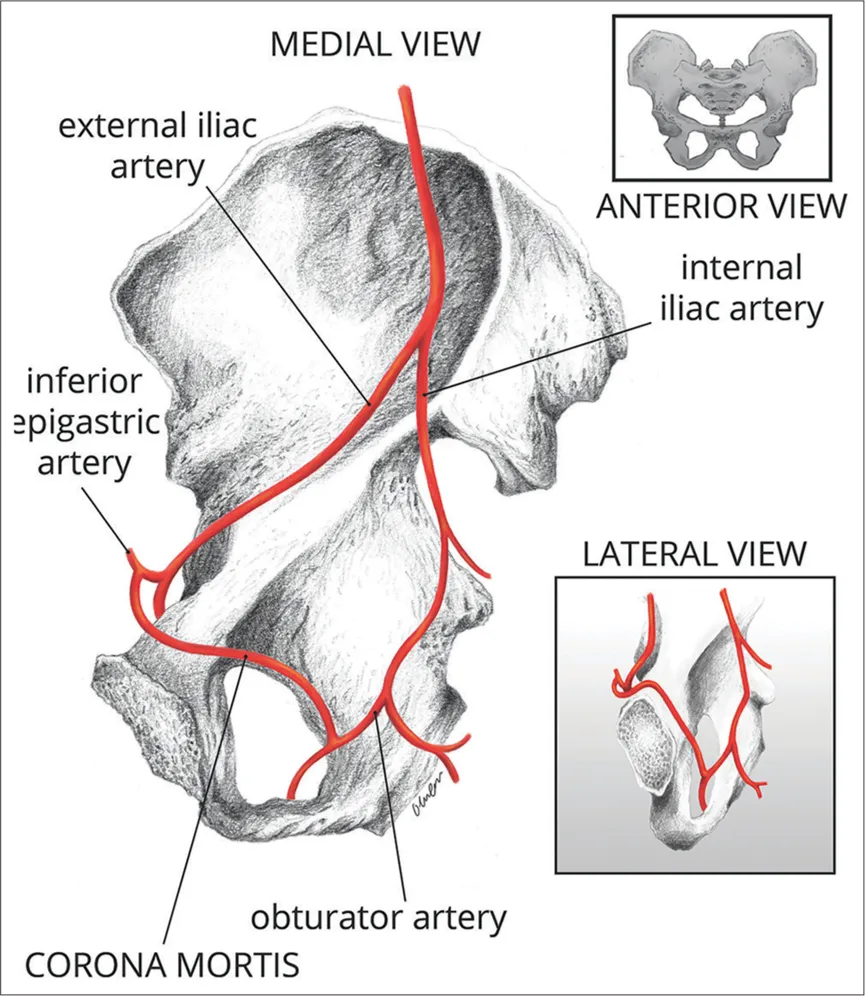
The Corona Mortis, ominously nicknamed the “Crown of Death,” is a vascular anomaly that poses significant risks in pelvic fracture surgeries. Its presence demands both anatomical awareness and precise hemorrhage control strategies to prevent life-threatening complications. This article delves into the anatomical significance, injury mechanisms, and evidence-based management techniques for this vascular variant, ensuring surgeons […]
Orthopedic Glossary: Essential Terms for Professionals
Understanding the language of orthopedics is fundamental for advancing expertise in the field. Whether you’re a surgeon, researcher, or student, mastering key terms enhances communication and comprehension of complex concepts. This glossary provides clear, concise definitions of essential orthopedic terminology, offering insights into procedures, materials, and pathologies. Dive into the world of orthopedic vocabulary and […]
Sports Medicine Research Over the Last Six Years: Everything You Need to Know
The field of sports medicine has undergone remarkable advancements in the past six years, with groundbreaking studies shaping how we diagnose, treat, and rehabilitate athletes. From ACL reconstruction techniques to emerging biologic therapies, this article dissects the latest trends, highlights key findings, and explores future directions in sports injury management. Whether you’re a clinician, researcher, or athlete, this comprehensive […]