

Meta Description:
This Part II analysis explains how orthopedic internal fixation failure occurs in long bone shaft fractures and unstable intertrochanteric fractures, and outlines anatomy-driven prevention strategies, technical details, and biomechanical principles to reduce fatigue failure and malunion.
(PAS Hook – Problem · Agitation · Solution)
Orthopedic internal fixation failure is rarely caused by poor implants. It is caused by misplaced confidence in technique. Surgeons often believe that once the biomechanical theory is understood, complications will naturally disappear. Reality is harsher. In long bone shaft comminution and unstable intertrochanteric fractures, the margin for error is almost zero. A single technical mistake—an entry point too lateral, ignored rotation, or a destroyed lateral wall—can doom fixation from the first postoperative step. Fatigue failure, deformity, and nonunion follow in predictable sequence. The solution is not stronger metal. It is anatomical precision, strain-aware strategy, and disciplined respect for biological limits.
(Featured Snippet – Direct Answer)
Orthopedic internal fixation failure in specific anatomical regions occurs when fixation strategies ignore local load-sharing requirements and biological tolerance. In long bone shaft comminuted fractures, unilateral plating fails when medial cortical support is absent, forcing plates to bear excessive axial and bending loads until fatigue fracture occurs. In unstable intertrochanteric fractures, failure results from misjudged instability—especially lateral wall disruption and rotational insufficiency—leading to uncontrolled collapse and malunion. Prevention relies on three principles: converting planar fixation into three-dimensional constructs, enhancing rotational stability without excessive biological damage, and preserving key anatomical structures such as the lateral wall through precise entry points and adjunct fixation techniques.
(Retainer – Transitional Paragraph)
Failure patterns repeat.
Anatomy does not change.
Only technique determines survival.
Orthopedic Internal Fixation Failure in Long Bone Shaft Comminuted Fractures
Why Orthopedic Internal Fixation Failure Occurs With Medial Cortical Defects
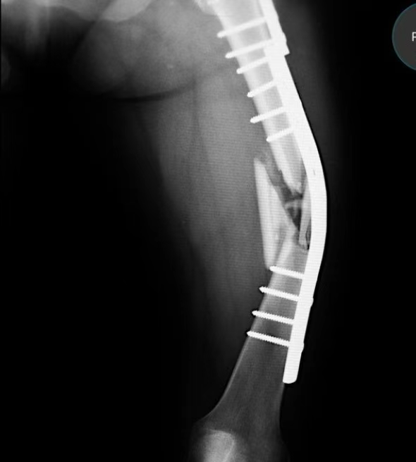
In long bone shaft comminuted fractures—particularly of the femur—the absence of medial cortical continuity creates a fatal mechanical imbalance. When a medial gap exists, lateral plates are forced to carry the entire axial and bending load. If fracture healing is delayed, the plate transitions from a load-sharing implant into a load-bearing beam. Fatigue fracture becomes inevitable. ICUC databases repeatedly confirm that this form of orthopedic internal fixation failure is mechanical in origin, not implant-related. Increasing plate thickness merely delays failure while worsening the biological environment through extensive soft-tissue stripping and contact-related osteoporosis. Once vascularity is compromised, even perfect mechanics cannot rescue healing.
Helical Plate Fixation as a Strategy to Prevent Orthopedic Internal Fixation Failure
The MIPO helical plate technique represents a strategic shift from mechanical confrontation to biomechanical cooperation. Instead of re-entering the original incision, a pre-contoured helical plate is slid through a distant minimal incision, wrapping partially around the bone. This plate intersects the primary fixation at approximately 90 degrees, converting a single-plane construct into a three-dimensional box-beam structure. The result is a dramatic increase in torsional and bending stiffness with minimal additional biological insult. Importantly, this approach often eliminates the need for bone grafting by restoring a favorable strain environment. Geometry—not bulk—becomes the solution to orthopedic internal fixation failure.
Plate Augmentation to Reduce Orthopedic Internal Fixation Failure After IM Nailing
For nonunion following intramedullary nailing, traditional teaching favors exchange nailing with aggressive reaming. While effective, this approach disrupts endosteal blood supply and places heavy physiological stress on the patient. ICUC data support plate augmentation as a less invasive alternative. By retaining the original nail and adding a unicortical locking plate or minimally invasive cerclage, surgeons selectively address rotational instability—the weakest aspect of IM fixation. This hybrid construct produces a qualitative mechanical upgrade while preserving biology. It is one of the most efficient ways to reverse orthopedic internal fixation failure without restarting the surgical trauma cycle.
Orthopedic Internal Fixation Failure in Unstable Intertrochanteric Fractures
Lateral Wall Failure as a Central Cause of Orthopedic Internal Fixation Failure
In intertrochanteric fractures, the lateral wall is the true gatekeeper of stability. Once compromised, neither DHS nor intramedullary nails can control collapse. Excessive telescoping follows, leading to limb shortening, abductor weakness, and persistent pain. Many failures originate from underestimating lateral wall fragility or causing iatrogenic blowout during reaming. Protecting the lateral wall is not optional—it is foundational. Loss of this structure remains one of the most common and preventable causes of orthopedic internal fixation failure in trochanteric fractures.
Rotational Instability and Orthopedic Internal Fixation Failure
Axial support alone does not guarantee success. During gait, the femoral head–neck fragment is subjected to repetitive rotational forces. Without adequate anti-rotation control, micro-motion accumulates, leading to reduction loss and implant migration. Dual-screw systems or DHS combined with trochanteric stabilizing plates address this weakness directly. Ignoring rotation is equivalent to ignoring physics. And physics, unlike surgeons, never negotiates.
Entry Point Errors That Trigger Orthopedic Internal Fixation Failure
A lateralized entry point is one of the most destructive technical errors in intertrochanteric fracture surgery. It directs reamers directly into the lateral wall, converting a manageable A2 fracture into an unstable A3 configuration. ICUC failure analyses repeatedly identify this mistake as a turning point from success to catastrophe. The correction is uncompromising: entry at the trochanteric apex or slightly medial, always with a protective sleeve. Minor injury to the gluteus medius tendon is acceptable. Lateral wall destruction is not.
Cerclage and Buttress Techniques to Prevent Orthopedic Internal Fixation Failure
The belief that cerclage “strangles bone” belongs to another era. Modern evidence from institutions such as HSS confirms that minimally invasive cerclage restores anatomy with negligible vascular compromise and substantial mechanical benefit (see: https://www.orthobullets.com). In long oblique or spiral subtrochanteric fractures, cerclage converts chaotic geometry into a stable tubular structure, allowing implants to function as intended. Anatomical reduction remains the strongest biological stimulus for healing. Without reduction, no fixation strategy can succeed.
Conclusion
Orthopedic internal fixation failure is not a hardware problem. It is an anatomical and strategic problem. In high-risk regions, survival depends on respecting local biomechanics, preserving biology, and executing technical details with absolute discipline.
Important Disclaimer:
- This article and all articles on this website are not popular science articles, but rather articles introducing advancements in medical research. All articles are for the learning and reference of medical professionals and for communication among those in the medical industry; specific medical issues should be addressed by consulting a doctor.
- Some articles on this website are generated with the assistance of AI and may contain inappropriate or even incorrect information. Please refer to the original English text if necessary, or leave feedback.
- We respect copyright. If your rights have been infringed, please contact us via WhatsApp at +8613915696091 to request removal.Thank you!
Like and share, your hands will be left with the fragrance!
More info. https://linktr.ee/shifreeman