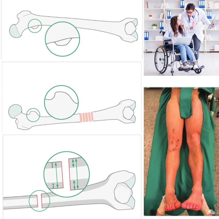
1. Assess the LT profile in comparison to the contralateral side
– Take a perfect lateral of the knee or an AP with the #patella centered over the distal femur then take an AP of the #hip
– the amount of visualized LT should be equal side to side post #femoral nailing
2. Key in fracture #fragments
3. Match #cortical thickness

4. Match the diameter difference between the proximal and distal fragment
5. Clinical comparison of contralateral leg rotation (hip IR and ER)
6. Utilize the anteversion built into the nail
– By centering the proximal locking screws in the femoral head and aligning the #distal locking #screws with the femoral condylar axis, the #proximal and distal fracture fragments are positioned in the same anteversion as the nail


